CT scans of visceral fat may help predict risk …C0NTINUE READING HERE >>>
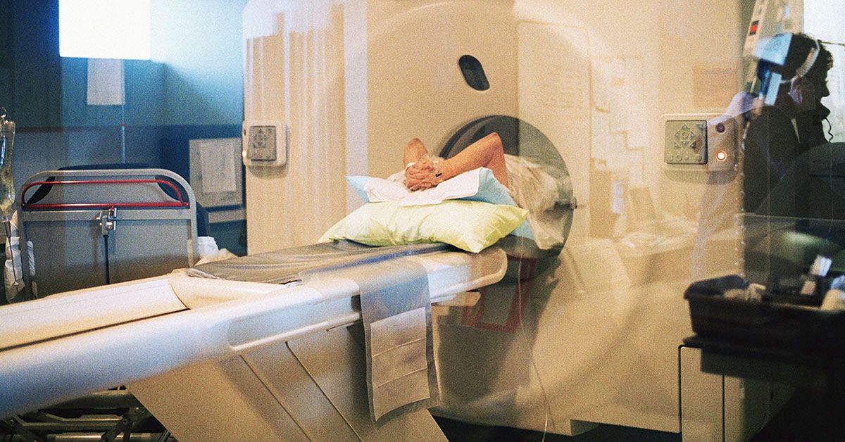
Share on PinterestResearchers are examining CT scans as potential tools to predict diabetes risk. Reza Estakhrian/Getty ImagesThere are many reasons why a person may be at an increased risk for type 2 diabetes. Having obesity or overweight and leading a sedentary lifestyle are some factors that can contribute to heightened risk.Researchers from Seoul, South Korea, have discovered that analyzing multi-organ CT scans can help doctors identify those who may have a heightened probability of type 2 diabetes.
About 90% to 95% of the estimated 529 million people worldwide living with diabetes have type 2 diabetes — a disease where a person doesn’t use insulin well, leading to blood sugar levels that are too high.
While some people may be genetically predisposed for type 2 diabetes, others increase their risk for the condition by following unhealthy lifestyle choices such as being overweight and inactive, as well as smoking and eating an unhealthy diet.
In an effort to help doctors better identify those who are at high risk for type 2 diabetes, researchers from Seoul, South Korea, have discovered that analyzing multi-organ computerized tomography (CT) scans can help doctors identify those who may have heightened probability.
The study was recently published in the journal Radiology.
For this study, researchers analyzed data from more than 32,000 adults with an average age of 45 who received a CT scan between January 2012 and December 2015.
“A CT scan is a medical imaging technique that uses X-rays to create detailed pictures of the inside of your body,” Seungho Ryu, MD, PhD, (he/him) professor of medicine at Kangbuk Samsung Hospital at Sungkyunkwan University School of Medicine in Seoul, South Korea and senior author of this study explained to Medical News Today.
“It takes many pictures from different angles, which are then combined to create cross-sectional images, like slices of bread, of the inside of your body. This allows doctors to see your bones, muscles, organs, and fat in much greater detail.”
“These detailed images can help identify the risk of type 2 diabetes by showing key indicators such as visceral fat (fat around internal organs), subcutaneous fat (fat under the skin), muscle mass and quality, liver fat, and aortic calcification (calcium build-up in the large arteries),” Ryu continued.
“High levels of visceral fat, poor muscle quality, and fatty liver are linked to a higher diabetes risk, while aortic calcification is associated with cardiovascular issues often seen in diabetes. This detailed information allows for early detection of who has diabetes and who may develop it, well before they become symptomatic and the condition becomes more serious.”
— Seungho Ryu, MD, PhD
Ryu and his team used deep learning algorithms to analyze study participants’ CT images. Using the CT markers of visceral fat, subcutaneous fat, muscle mass, liver density, and aortic calcium, researchers were able to determine a person’s type 2 diabetes risk.
Scientists found that the amount of visceral fat was the highest predictor of diabetes risk. Combining visceral fat with muscle area, liver fat fraction, and aortic calcification further improved diabetes predictions.
“Visceral fat and liver fat are known to significantly increase the risk of diabetes due to their roles in insulin resistance, a key mechanism of type 2 diabetes,” Yoosoo Chang, MD, PhD, professor of medicine at Kangbuk Samsung Hospital at Sungkyunkwan University School of Medicine in Seoul, South Korea, and co-first author of this study, explained to MNT.
“Skeletal muscle mass and quality, which regulate glucose homeostasis and are essential for metabolism, exercise, and metabolic disease management, can be assessed using CT images. Aortic calcification serves as a cumulative marker of cardiovascular risk over a lifetime and is recently considered a general aging marker beyond cardiovascular risk.”
— Yoosoo Chang, MD, PhD
“Fortunately, all these measures are easily obtained from a fully automated AI solution,” Chang added. “Combining these markers provides a comprehensive picture of an individual’s metabolic state, enhancing the accuracy of diabetes risk prediction.”
At the start of the study, diabetes frequency was 6%, and occurrence rose to 9% during the average 7.3-year follow-up period.
“Based on the reported prevalence of diabetes in 2016 — 13.7% among Korean adults aged 30 years and above — the subjects in this study were at a relatively low risk of diabetes,” Soon Ho Yoon, MD, PhD, professor of medicine at Seoul National University Hospital, Seoul National College of Medicine in Seoul, South Korea, and co-first author of this study, told MNT.
“Despite the study subjects’ lower risk, the predictive power of body composition analysis highlights its potential utility in identifying individuals at risk of developing diabetes,” he said.
“As the prevalence of diabetes continues to grow globally, leveraging previously unused imaging information for early detection and risk identification can significantly enhance preventive efforts and patient outcomes,” Yoon added.
“As the next steps for this research, we plan to validate our findings in diverse populations, particularly in the U.S.,” Ryu said. “This will involve collaborating with competitive U.S. researchers who are also investigating the adjunctive role of advanced medical imaging tools. Additionally, assessing the cost-effectiveness of using CT scans for opportunistic screening is mandatory. Exploring other CT-derived markers to enhance predictive accuracy and evaluate additional diseases will also be a focus.”
“We utilized organ quantification information available at the start of the study, but other organs, such as the pancreas, kidneys, and other chest organs, are becoming available for analysis,” he continued.
“We hope to improve the performance of the prediction model for various major chronic diseases, including diabetes and other cardiometabolic diseases, by incorporating additional image quantification data. Ultimately, we hope to assess how early identification and intervention based on CT-derived markers influence patient outcomes by integrating these advanced imaging techniques into routine clinical practice.”
— Seungho Ryu, MD, PhD
MNT also spoke with Pouya Shafipour, MD, a board certified family and obesity medicine physician at Providence Saint John’s Health Center in Santa Monica, CA, about this study, who said he was not surprised by its results.
“Abdominal adiposity is one of the highest risks for development of diabetes (and) prediabetes,” Shafipour explained. “Fatty liver and insulin resistance are usually the prodromes to onset of [d]iabetes, so I was not surprised.”
“Diabetes, prediabetes, and insulin resistance is a continuum, so when someone is diagnosed with diabetes, they’re often in this prediabetic state for a long period of time. The earlier we can detect this, the earlier we can take action, we can start coaching the patient, the less costly, and the more effective and reversible it will be. So the CT scan seems to be catching it earlier than some of the other conventional models.”
— Pouya Shafipour, MD
Shafipour commented that potential downsides to using CT scans may be the cost and risk of radiation.
“Patients get CT scans for different reasons,” he explained.
“If this can be put into some type of guideline or calculation so your traditional radiologists when they’re reading regular CT scans can be like, okay, … this is where they stand in terms of visceral fat and potential risk of diabetes so it can guide whoever has ordered that CT scan to take action to either treat them themselves, make referrals for dieticians, counseling, specialists, obesity medicine, things like that,” he added. “I think that would be very helpful.”
>
